PRODUCTS SOLD ON PEPTIDESLABEU.COM ARE FOR RESEARCH PURPOSES ONLY AND ARE NOT FOR HUMAN OR VETERINARY USE.
€207.00
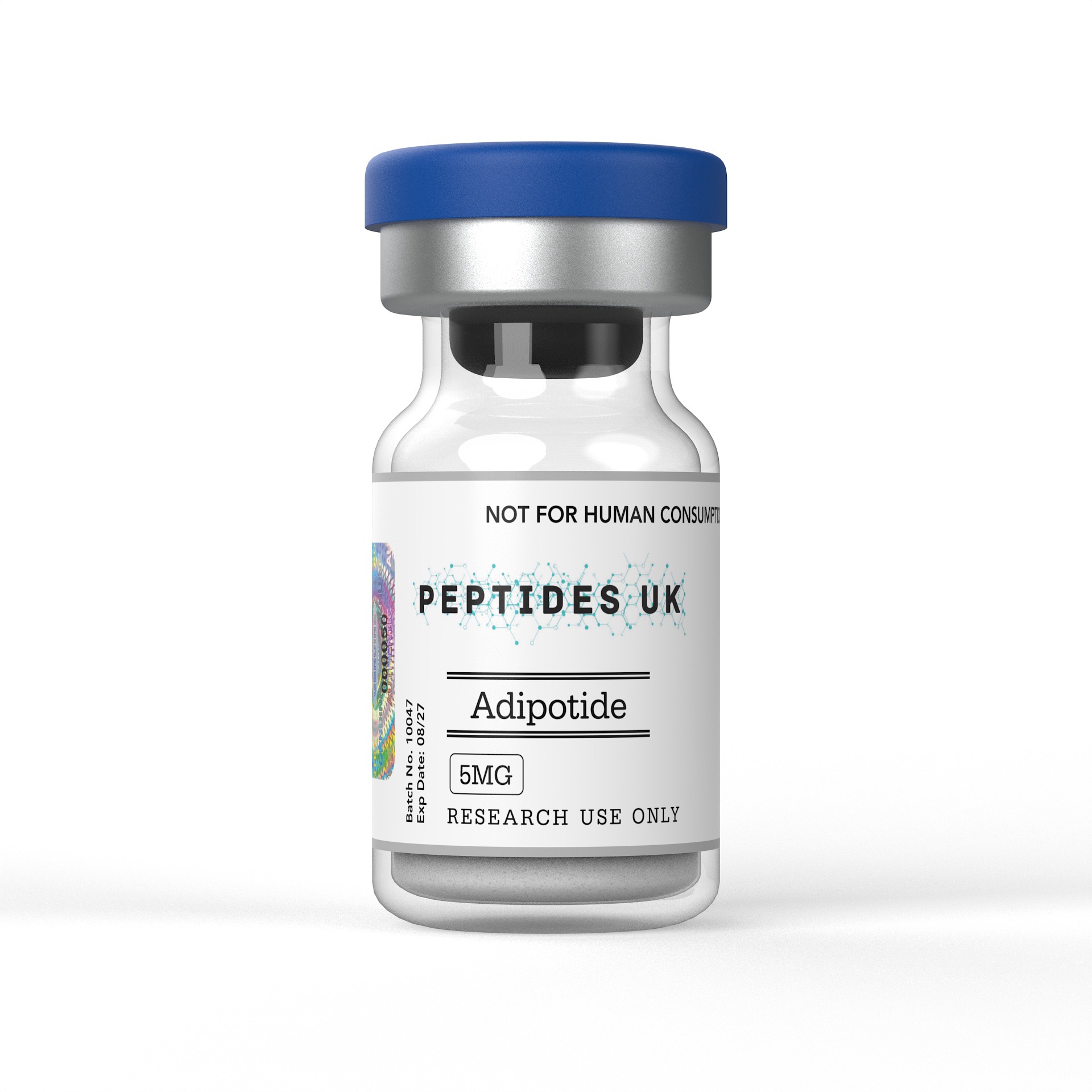
Adipotide EU – Buy Online | In Stock & Ready to Ship
Buy Adipotide in Europe with fast shipping and guaranteed ≥99% purity — verified with COA and HPLC documentation. A trusted choice for peptides EU research teams rely on, with no customs delays or lengthy international wait times. Whether you’re searching for Adipotide Europe suppliers, looking to buy Adipotide in the EU, or sourcing peptides Europe-wide, we have you covered. Research teams across the EU can count on consistent stock, rapid fulfilment and full batch documentation every time.
For research use only. Not intended for human or veterinary use.

Adipotide is a synthetic pro-apoptotic peptidomimetic — a chimeric bimodal peptide constructed by fusing a white adipose tissue vasculature-homing domain (CKGGRAKDC) to a pro-apoptotic mitochondrial membrane-disrupting sequence (D(KLAKLAK)₂) through a GG dipeptide linker — that selectively targets and destroys the blood vessel endothelium supplying white adipose tissue (WAT), inducing apoptosis of adipose-resident vasculature, consequent ischaemic death of WAT adipocytes dependent on that vasculature for survival, and profound fat mass reduction in preclinical models, available to buy in Europe for laboratory research into adipose tissue vascular biology, WAT-targeted apoptotic strategies, prohibitin-mediated adipose vasculature homing, body composition regulation, pro-apoptotic peptide pharmacology, vascular-targeted obesity biology, and the comparative study of adipose tissue-selective cytotoxic approaches.
Laboratories and research institutions across the EU can order verified, research-grade adipotide with fast international dispatch to Europe, full batch documentation, and ≥99% purity confirmed by HPLC and Mass Spectrometry.
✅ ≥99% Purity — HPLC & Mass Spectrometry Verified
✅ Batch-Specific Certificate of Analysis (CoA)
✅ Sterile Lyophilised Powder | GMP Manufactured
✅ Fast Dispatch to EU & Europe | Tracked Shipping
Adipotide (also designated CKGGRAKDC-GG-D(KLAKLAK)₂, or in earlier literature as PROHIBITIN-TARGETED PROAPOPTOTIC PEPTIDE or fat-targeted peptidomimetic) is a synthetic chimeric peptide developed by Wadih Arap, Renata Pasqualini, and colleagues at The University of Texas MD Anderson Cancer Center, arising from a programme of phage display-based in vivo peptide library screening designed to identify vascular zip code sequences — short peptide motifs that home selectively to the blood vessel endothelium of specific tissues and organs when administered systemically — that could serve as targeting vectors for the delivery of cytotoxic payloads to defined tissue vasculatures.
The CKGGRAKDC sequence — adipotide’s N-terminal homing domain — was identified through in vivo phage display screening in mice and subsequently in non-human primates as a peptide motif that homes selectively to the endothelium of white adipose tissue vasculature when injected systemically, binding a receptor complex at the surface of WAT endothelial cells while exhibiting markedly lower binding to the vasculature of other tissues including skeletal muscle, heart, liver, kidney, and brain. Subsequent molecular studies identified prohibitin — a multifunctional protein with well-characterised roles in mitochondrial inner membrane architecture and electron transport chain complex assembly that also appears, unexpectedly, at the surface of white adipose tissue endothelial cells — as the molecular receptor mediating CKGGRAKDC’s selective WAT vascular homing. The cell-surface localisation of prohibitin on WAT endothelium — a localisation absent or markedly lower in endothelial cells of other tissues — constitutes the molecular specificity determinant that underlies adipotide’s tissue selectivity.
The D(KLAKLAK)₂ sequence — adipotide’s C-terminal pro-apoptotic effector domain — is a D-amino acid analogue of the (KLAKLAK)₂ amphipathic lytic peptide originally developed as an antibacterial peptide that disrupts bacterial membranes. In the context of intracellular delivery to mammalian cells, D(KLAKLAK)₂ disrupts mitochondrial inner membranes — the double-membrane organelle compartment where electron transport and oxidative phosphorylation occur — through an amphipathic mechanism involving electrostatic interaction with negatively charged mitochondrial membrane phospholipids and hydrophobic membrane insertion that permeabilises the mitochondrial inner membrane, collapses the mitochondrial membrane potential (ΔΨm), releases cytochrome c, and triggers the intrinsic apoptotic pathway. D-amino acid stereochemistry confers resistance to cellular proteases, ensuring that the effector domain remains intact following endosomal escape and cytoplasmic delivery. The GG dipeptide linker connecting the homing and effector domains provides structural flexibility enabling each domain to adopt its functional conformation independently.
The mechanistic logic of adipotide is therefore vascular-targeted: systemic administration delivers the intact chimeric peptide to the circulation, where CKGGRAKDC-mediated prohibitin binding selectively tethers adipotide to WAT endothelial cell surfaces, internalisation delivers D(KLAKLAK)₂ to the intracellular compartment, mitochondrial membrane disruption drives apoptotic death of WAT endothelial cells, the consequent loss of vascular supply produces ischaemia and apoptotic-necrotic death of the adipocytes and stromal cells that depend on that vasculature, and the net result is a selective reduction in white adipose tissue mass without direct cytotoxic effects on tissues whose vasculature lacks surface prohibitin expression at levels sufficient for adipotide accumulation.
In laboratory settings, adipotide is studied across adipose tissue vascular biology, prohibitin surface biology in WAT endothelium, pro-apoptotic peptide delivery mechanisms, body composition and obesity pharmacology, adipose tissue ischaemia and cell death biology, vascular zip code targeting methodology, and the comparative biology of WAT versus other adipose depot responses to vascular-targeted interventions. EU and European researchers working with adipotide typically focus on:
White adipose tissue vascular homing and prohibitin surface biology research — The most mechanistically foundational research application of adipotide is as a molecular probe for prohibitin surface expression in WAT endothelium — using fluorescently labelled, biotinylated, or otherwise tagged adipotide constructs to map the distribution and density of surface prohibitin across endothelial cell populations from different tissue vascular beds, characterise the molecular determinants of prohibitin’s atypical cell-surface localisation in WAT versus its canonical intracellular mitochondrial and nuclear localisations, and establish the structural requirements of the CKGGRAKDC/prohibitin interaction using alanine scanning mutagenesis and competitive inhibition studies. These studies address a fundamental cell biology question — how and why prohibitin reaches the outer leaflet of WAT endothelial plasma membranes — that has implications for understanding adipose tissue vascular identity and for identifying additional WAT endothelial surface markers exploitable for targeted delivery.
Pro-apoptotic peptide intracellular delivery and mitochondrial disruption research — The D(KLAKLAK)₂ effector domain of adipotide is a well-established pro-apoptotic mitochondrial membrane-disrupting sequence that has been extensively studied in the context of targeted delivery constructs. Studies characterising adipotide’s apoptotic mechanism in WAT endothelial cells examine the intracellular trafficking of adipotide following prohibitin-mediated surface binding and internalisation — characterising endosomal escape efficiency, cytoplasmic distribution, mitochondrial co-localisation (using MitoTracker, TOM20 co-staining), mitochondrial membrane potential dissipation (JC-1, TMRE fluorescent probes), cytochrome c release from mitochondria to cytoplasm, caspase-9 and caspase-3/7 activation downstream of cytochrome c-driven apoptosome formation, and the morphological features of intrinsic apoptosis in adipotide-treated endothelial cells — establishing the complete apoptotic signalling cascade from prohibitin surface binding to mitochondrial outer membrane permeabilisation and caspase-dependent cell death execution.
Adipose tissue vascular biology and endothelial heterogeneity research — Adipotide’s WAT endothelial selectivity provides a functional tool for characterising the molecular heterogeneity of endothelial cells across different tissue vascular beds — specifically the features that distinguish WAT endothelium from muscle, liver, cardiac, renal, and brain endothelium at the level of surface proteome composition. Studies use adipotide binding, combined with proteomic characterisation of WAT endothelial surface-accessible proteins (surface biotinylation proteomics, proximity labelling), to build a molecular portrait of WAT vascular endothelial identity — identifying additional WAT endothelial surface markers co-expressed with prohibitin and establishing adipose tissue vascular zip codes as a molecular framework for understanding organ-specific vascular specialisation.
Body composition and adipose tissue mass regulation research — In diet-induced obese rodent and non-human primate models, adipotide treatment produces rapid, substantial reductions in white adipose tissue mass — measured by MRI-based body composition analysis, adipose depot weighing, adipocyte size quantification, and whole-body fat percentage assessment — through WAT vascular apoptosis and consequent adipocyte ischaemic death. Studies characterise the dose-response relationship between adipotide administration and WAT mass reduction, the anatomical specificity of fat depot targeting (subcutaneous versus visceral WAT depots), the time course of WAT involution following vascular apoptosis, and the reversibility or permanence of adipose tissue loss after adipotide treatment — establishing the pharmacological parameters of WAT-targeted vascular apoptosis as a fat mass reduction strategy.
Adipose depot selectivity — visceral versus subcutaneous WAT biology — White adipose tissue is anatomically and functionally heterogeneous — visceral adipose depots (omental, mesenteric, retroperitoneal WAT) differ metabolically and in their cardiovascular risk associations from subcutaneous adipose depots (inguinal, dorsal, femoral WAT) — and understanding whether adipotide acts equivalently across these depots, or shows differential activity reflecting potential differences in prohibitin surface expression levels between visceral and subcutaneous WAT vasculature, is a mechanistically and translationally relevant research question. Studies use adipotide in models with quantifiable depot-specific fat mass measurements — including MRI-based visceral/subcutaneous fat volumetry in rodents and primates — to characterise the relative sensitivity of different WAT depots to adipotide-induced vascular apoptosis and to determine whether depot-specific differences in WAT endothelial prohibitin expression contribute to differential adipotide sensitivity.
Brown adipose tissue and beige adipose tissue selectivity research — Brown adipose tissue (BAT) and beige/brite adipocytes within white fat depots are thermogenically active fat cells with distinct vascular supply characteristics, endothelial gene expression profiles, and potentially different prohibitin surface expression levels compared to WAT endothelium. Studies examining adipotide’s selectivity between WAT and BAT vasculature — using adipotide binding assays in primary BAT versus WAT endothelial cell isolates, fluorescent adipotide biodistribution in animals with defined BAT depots, and BAT-specific histology and UCP1 immunostaining post-adipotide treatment — characterise whether adipotide’s WAT endothelial targeting extends to BAT vasculature and whether BAT thermogenic function is preserved or impaired following adipotide-induced fat depot remodelling.
Metabolic consequences of WAT mass reduction research — Adipose tissue is not merely an energy storage depot but a highly active endocrine organ secreting adipokines (leptin, adiponectin, resistin, TNF-α, IL-6) that regulate systemic insulin sensitivity, energy balance, and inflammation. Studies examining the metabolic consequences of adipotide-induced WAT mass reduction characterise adipokine secretion profiles following adipotide treatment — including changes in circulating leptin, adiponectin, and pro-inflammatory adipokines — and assess systemic metabolic parameters including insulin sensitivity (glucose and insulin tolerance tests, euglycaemic-hyperinsulinaemic clamp), hepatic steatosis (reflecting ectopic lipid redistribution following peripheral fat depot loss), and inflammatory marker profiles — establishing the systemic metabolic phenotype associated with pharmacological WAT vascular apoptosis.
Kidney function and nephrotoxicity biology in vascular-targeted peptide research — Preclinical adipotide studies in obese non-human primates documented evidence of nephrotoxic effects — including elevations in blood urea nitrogen (BUN) and creatinine, and histological evidence of renal tubular injury — that were transient and reversible with treatment cessation but that represent a mechanistically important research finding regarding the off-target tissue consequences of WAT vascular-targeted apoptosis at pharmacologically active doses. Studies examining adipotide’s renal effects characterise the mechanism of nephrotoxicity — whether reflecting direct prohibitin-mediated effects on renal tubular cells (which express prohibitin intracellularly and potentially at low surface density), haemodynamic changes secondary to rapid fat loss, or altered renal handling of apoptotic cellular debris from WAT involution — and identify dose and schedule parameters that dissociate WAT-targeted fat loss from renal toxicity.
Vascular zip code peptide methodology and in vivo phage display research — The CKGGRAKDC homing peptide was identified using in vivo phage display — a methodology in which bacteriophage libraries displaying diverse peptide sequences are injected systemically into living animals, the phage population that homes to a specific tissue vasculature is recovered and amplified through iterative biopanning rounds, and individual tissue-homing sequences are identified by sequencing phage inserts from the enriched tissue-specific population. Adipotide is used as a validated positive control and proof-of-concept example in studies establishing and optimising in vivo phage display methodologies — demonstrating the identification, validation, and functional exploitation of vascular zip code sequences — and in comparative studies examining the degree of conservation of WAT vascular homing sequences between rodent, non-human primate, and human adipose tissue vasculature.
Combination with conventional obesity treatments and metabolic intervention research — Studies examining adipotide in combination with dietary restriction, exercise interventions, GLP-1 receptor agonists, or other pharmacological obesity treatments characterise whether WAT vascular apoptosis and pharmacological approaches to obesity engage complementary or overlapping mechanisms — providing mechanistic insight into whether fat mass reduction through ischaemic adipocyte death produces different metabolic signatures from fat mass reduction through energy deficit-driven adipocyte lipid mobilisation, and whether the combination of vascular-targeted WAT apoptosis with metabolic pharmacology produces additive or synergistic body composition changes.
Prohibitin biology — multifunctional protein research — Prohibitin (PHB1) is a protein of remarkable functional versatility — well characterised as a structural component of the mitochondrial inner membrane where it forms ring-like PHB1/PHB2 heterodimeric complexes that scaffold the assembly of respiratory chain complexes and regulate mitochondrial morphology and dynamics; also present in the nucleus where it modulates transcription factor activity (including E2F, p53, and Rb interactions); and, as established through adipotide’s homing biology, expressed at the surface of WAT endothelial cells as the receptor for CKGGRAKDC. Studies use adipotide as a surface prohibitin probe to investigate the biology of prohibitin’s unconventional cell-surface localisation — characterising the mechanism of prohibitin trafficking from its mitochondrial and nuclear compartments to the plasma membrane outer leaflet in WAT endothelial cells, the post-translational modifications or binding partners governing this trafficking, and the functional significance of surface prohibitin for WAT endothelial cell biology independent of adipotide’s apoptotic payload.
All research applications are for in vitro and pre-clinical use only.
Adipotide has a foundational but influential research literature — anchored in the landmark papers by Kolonin, Saha, Chan, Pasqualini, and Arap that established WAT vascular targeting through in vivo phage display, the prohibitin surface receptor identification, and the preclinical efficacy in rodent and non-human primate models — with subsequent studies extending the biology to metabolic consequences, mechanism characterisation, and comparative fat depot analysis.
Foundational in vivo phage display identification and WAT vascular homing characterisation: The landmark studies establishing adipotide’s biology employed iterative in vivo phage display biopanning in mice — systematically enriching for phage clones homing to adipose tissue vasculature versus other organs — and identified CKGGRAKDC as a sequence displaying selective homing to white adipose tissue endothelium detectable by phage recovery quantification, histological co-localisation of phage particles with WAT blood vessels, and fluorescent peptide biodistribution studies showing preferential accumulation of CKGGRAKDC-displaying peptides in WAT versus muscle, liver, heart, kidney, and brain vasculature. The selectivity of homing was validated across multiple WAT depots (subcutaneous inguinal, epididymal visceral, omental) and confirmed in independent experiments using synthetic CKGGRAKDC peptide rather than phage-displayed constructs.
Prohibitin surface receptor identification: Studies characterising the molecular receptor for CKGGRAKDC homing used affinity chromatography with immobilised CKGGRAKDC peptide coupled to mass spectrometric protein identification from WAT endothelial cell lysates — identifying prohibitin as the primary CKGGRAKDC-binding protein in WAT endothelial cells. Surface biotinylation proteomics and flow cytometry with anti-prohibitin antibodies on non-permeabilised WAT endothelial cells confirmed cell-surface localisation of prohibitin in WAT endothelium — a localisation not previously described for this classically intracellular protein and not detected at equivalent levels on endothelial cells from other tissues. Competitive inhibition of CKGGRAKDC binding by anti-prohibitin antibodies, and loss of adipotide homing efficacy in prohibitin-knockdown WAT endothelial cells, established the causal role of surface prohibitin in adipotide’s tissue-selective targeting.
Obese mouse model fat reduction studies: Studies in genetically obese (ob/ob) and diet-induced obese mice treated with adipotide documented rapid, dose-dependent reductions in white adipose tissue mass — with treated animals losing substantial body weight driven by fat mass loss rather than lean mass reduction, as confirmed by MRI-based body composition analysis. Histological examination of WAT depots from adipotide-treated animals revealed endothelial cell apoptosis (TUNEL positivity, activated caspase-3 immunostaining in WAT blood vessels), vascular regression, adipocyte shrinkage and apoptosis, and adipose tissue involution — establishing the causal chain from WAT vascular apoptosis to adipose tissue mass reduction. Pair-housing and pair-feeding controls confirmed that the body weight loss was not secondary to adipotide-induced food intake suppression or behavioural effects.
Non-human primate obesity model studies: Extending the rodent findings, studies in obese rhesus macaques treated with adipotide documented significant reductions in body weight and MRI-quantified body fat percentage over the treatment period — with visceral fat showing particularly pronounced reductions. Metabolic profiling in treated primates demonstrated improvements in insulin sensitivity indices accompanying fat mass reduction. These non-human primate studies — representing the closest preclinical model to human physiology — were mechanistically significant in confirming that the WAT vascular homing biology of CKGGRAKDC was conserved between rodent and primate adipose vasculature and that pharmacologically meaningful fat reduction was achievable through this mechanism in a primate model. The same studies documented the reversible nephrotoxicity findings at higher dose levels, establishing the translational significance of the renal safety consideration.
Mechanistic apoptosis pathway characterisation: Studies characterising the apoptotic mechanism in adipotide-treated WAT endothelial cells confirmed the intrinsic (mitochondrial) apoptosis pathway — documenting mitochondrial membrane potential collapse (JC-1 assay), cytochrome c cytoplasmic translocation, caspase-9 activation upstream of caspase-3/7 effector caspase activation, and the absence of significant caspase-8 activation (extrinsic pathway) — establishing D(KLAKLAK)₂-mediated mitochondrial membrane disruption as the apoptotic initiation event following prohibitin-directed intracellular delivery in WAT endothelial cells.
Prohibitin functional biology studies: Studies examining prohibitin’s role in WAT endothelial cell biology — prompted by its identification as adipotide’s surface receptor — characterised PHB1 expression patterns across endothelial cells from different tissue origins, the trafficking pathway by which prohibitin reaches the WAT endothelial cell surface, and the functional consequences of prohibitin knockdown on WAT endothelial cell viability, angiogenic capacity, and adipose tissue vascularisation — establishing prohibitin as a functional component of WAT endothelial biology with a role beyond its canonical mitochondrial and nuclear functions, and identifying the WAT endothelial surface prohibitin axis as a molecular feature distinguishing adipose vasculature from the endothelium of other organs.
Comparative tissue selectivity and normal tissue sparing studies: Studies examining adipotide’s effects on non-adipose tissues — including skeletal muscle, cardiac muscle, liver, kidney (at non-nephrotoxic doses), brain, and bone marrow — documented the absence of significant apoptosis, vascular regression, or tissue weight reduction in these organs at doses producing substantial WAT mass reduction, consistent with the low-level or absent prohibitin surface expression in the vasculature of these tissues. This selectivity characterisation established the tissue-targeting index of adipotide and provided the mechanistic basis for its potential utility as a fat-selective cytotoxic approach compared to systemic metabolic or pharmacological interventions that affect multiple tissue types.
| Compound | Class | Targeting Mechanism | Effector Mechanism | Target Cell | Selectivity Determinant | Key Research Distinction |
|---|---|---|---|---|---|---|
| Adipotide (CKGGRAKDC-GG-D(KLAKLAK)₂) | Chimeric pro-apoptotic peptidomimetic | CKGGRAKDC → surface prohibitin on WAT endothelium | D(KLAKLAK)₂ → mitochondrial membrane disruption → intrinsic apoptosis | WAT blood vessel endothelial cells | Surface prohibitin expression on WAT endothelium | WAT vascular-targeted; prohibitin surface biology probe; intrinsic apoptosis via mitochondrial disruption; fat mass reduction through ischaemic adipocyte death |
| D(KLAKLAK)₂ alone | Pro-apoptotic mitochondrial lytic peptide (untargeted) | None — non-selective cellular uptake | Mitochondrial membrane disruption → intrinsic apoptosis | Non-selective (any cell with uptake) | None | Essential control — effector domain without WAT homing; establishes prohibitin-dependent targeting contribution to adipotide selectivity |
| CKGGRAKDC alone | WAT endothelial homing peptide (no effector) | Surface prohibitin binding — WAT endothelium | None | WAT endothelial surface binding only | Surface prohibitin | Homing domain control; competitive inhibitor of adipotide; surface prohibitin probe without cytotoxic payload |
| RGD-D(KLAKLAK)₂ | Integrin-targeted pro-apoptotic construct | RGD → αvβ3/αvβ5 integrin — tumour neovasculature | D(KLAKLAK)₂ → mitochondrial disruption | Tumour neovasculature endothelium | αvβ3/αvβ5 integrin overexpression | Tumour vasculature comparator; same effector domain; different homing peptide; establishes transferability of D(KLAKLAK)₂ delivery strategy |
| NGR-D(KLAKLAK)₂ | Aminopeptidase N-targeted pro-apoptotic construct | NGR → aminopeptidase N (CD13) — tumour vasculature | D(KLAKLAK)₂ → mitochondrial disruption | Tumour neovasculature | CD13/aminopeptidase N | Tumour vasculature comparator; vascular zip code targeting strategy with same effector; established in tumour angiogenesis research |
| Prohibitin (PHB1) antibodies | Prohibitin-targeting research tool | Direct anti-PHB1 binding — surface prohibitin | No cytotoxic payload | WAT endothelium surface | Surface prohibitin | Surface prohibitin localisation probe; competitive inhibitor of adipotide binding; establishes prohibitin-dependence of adipotide homing |
| Bevacizumab / anti-VEGF | Anti-angiogenic antibody | VEGF neutralisation — blocks VEGFR2 signalling | Angiogenesis inhibition (anti-proliferative, not apoptotic) | Endothelial cells — VEGF-dependent angiogenesis | VEGF overexpression in tumour/tissue | Mechanistic comparator — anti-angiogenic vs vascular-apoptotic; non-selective endothelial targeting vs WAT-specific; anti-tumour angiogenesis context |
Every order of adipotide dispatched to EU and European research institutions includes:
Can I Buy Adipotide in the EU and Europe?
Yes. We supply research-grade adipotide with fast tracked dispatch to all EU member states and wider European destinations. All orders include full batch documentation. Adipotide is supplied strictly for laboratory research use only.
What is the Mechanistic Logic of the Bimodal Chimeric Peptide Design and Why Are Both Domains Essential?
Adipotide’s activity depends on the obligate functional cooperation of both domains within the intact chimeric peptide. The CKGGRAKDC homing domain provides WAT endothelial specificity through surface prohibitin binding — delivering the intact construct to the plasma membrane of WAT endothelial cells while the peptide circulates through other tissues without significant accumulation. Without the homing domain, D(KLAKLAK)₂ alone is taken up non-selectively by all cells proportional to their endocytic activity — producing non-selective cytotoxicity that is not WAT-specific. Conversely, CKGGRAKDC without the D(KLAKLAK)₂ effector domain binds WAT endothelial surface prohibitin but produces no apoptotic or cytotoxic consequence. The chimeric design achieves selectivity through two sequential specificity filters: the first at the level of WAT endothelial surface binding (prohibitin-dependent tissue homing), and the second at the intracellular level (mitochondrial membrane disruption requires successful internalisation and endosomal escape — steps that are substantially more efficient in cells where the construct is concentrated at the surface by high-affinity receptor binding than in cells where uptake is purely non-specific). The GG dipeptide linker connecting the domains provides conformational flexibility — its function has been confirmed by studies showing that direct fusion without a linker reduces activity, whereas the diglycine spacer preserves the independent folding and function of each domain.
Why is Prohibitin Found at the Surface of WAT Endothelial Cells But Not Other Endothelium?
The molecular mechanism governing prohibitin’s selective surface exposure in WAT endothelium — compared to its predominantly intracellular mitochondrial and nuclear localisation in most other cell types — remains an active area of investigation and an incompletely resolved research question. Current evidence suggests that WAT endothelial cells undergo an active trafficking process delivering a fraction of prohibitin to the plasma membrane outer leaflet — potentially involving unconventional secretion pathways, membrane microvesicle shedding and reincorporation, or lipid raft-associated trafficking — that is regulated by signals specific to the WAT tissue microenvironment. Candidate regulatory factors include adipocyte-derived signals (fatty acids, adipokines such as adiponectin, VEGF isoforms produced by adipocytes) that programme WAT endothelial cell identity and may drive prohibitin surface trafficking as part of a broader WAT endothelial transcriptional and proteomic programme. Post-translational modifications of prohibitin — including phosphorylation at Thr²⁵⁸, Tyr²⁵⁹, and other residues — have been identified as potential regulators of its subcellular distribution. The tissue-specific surface prohibitin phenotype of WAT endothelium represents a fundamental cell biology question with implications for adipose vascular identity, and adipotide’s CKGGRAKDC homing domain serves as a molecular tool for probing this biology.
How Does Adipotide’s Mechanism Differ From GLP-1 Receptor Agonists and Other Pharmacological Obesity Treatments?
Adipotide and pharmacological obesity treatments — including GLP-1 receptor agonists (semaglutide, liraglutide), amylin analogues (cagrilintide), and other centrally acting appetite suppressants — reduce body fat mass through fundamentally different mechanisms that operate at entirely different biological levels. GLP-1 receptor agonists and amylin analogues act centrally through hypothalamic and brainstem satiety circuits to reduce food intake — producing a negative energy balance in which the organism consumes less fuel than it expends, driving mobilisation of stored triglycerides from adipocytes and consequent reduction in adipocyte lipid content and adipose mass. In this process, the adipose vasculature is preserved; adipocytes shrink through lipid mobilisation but remain viable, and fat mass reduction is in principle reversible if energy balance is restored. Adipotide acts peripherally and directly on adipose tissue vasculature — destroying the blood supply to white fat depots and producing ischaemic cell death of the dependent adipocytes regardless of energy balance or food intake. The fat cell death mechanism (ischaemia-driven necrosis and secondary apoptosis) is more destructive and potentially less reversible than the lipid mobilisation mechanism of appetite-suppressing agents. For research, this mechanistic distinction enables controlled comparisons of the metabolic consequences of fat mass reduction through energy deficit versus adipocyte cell death — with potential differences in adipokine profiles, adipose inflammation, hepatic lipid redistribution, and long-term adipose tissue remodelling.
What Are the Key Controls Required for Mechanistic Adipotide Research?
Rigorous mechanistic interpretation of adipotide experiments requires several layers of controls addressing both the homing and effector components. CKGGRAKDC alone (homing peptide without effector) establishes the baseline effect of surface prohibitin binding without apoptotic payload — confirming that WAT endothelial surface binding per se does not produce cytotoxicity and that the observed apoptotic effects require D(KLAKLAK)₂ delivery. D(KLAKLAK)₂ alone (effector without homing domain) establishes the non-selective baseline toxicity of the pro-apoptotic domain in the absence of WAT-specific targeting — confirming that adipotide’s selectivity requires CKGGRAKDC-mediated tissue targeting. A scrambled CKGGRAKDC sequence fused to D(KLAKLAK)₂ (sequence-scrambled adipotide with identical amino acid composition) confirms that the CKGGRAKDC sequence — not its composition — drives WAT homing, and that the observed WAT selectivity is not an artefact of the peptide’s overall charge or hydrophobicity. Anti-prohibitin antibody pre-treatment of WAT endothelial cells prior to adipotide addition — blocking surface prohibitin — should attenuate adipotide binding and cytotoxicity proportionally, confirming prohibitin-dependence. Prohibitin siRNA knockdown in WAT endothelial cells should similarly reduce adipotide sensitivity, establishing the causal role of surface prohibitin. For in vivo fat mass studies, MRI-based body composition analysis distinguishing fat mass from lean mass is essential to confirm that weight loss reflects adipose tissue loss rather than lean tissue wasting.
Is Adipotide Selective for Visceral or Subcutaneous White Adipose Tissue?
Preclinical studies have documented adipotide activity across multiple WAT depots — including both visceral (omental, mesenteric, epididymal) and subcutaneous (inguinal, dorsal) depots — consistent with the hypothesis that surface prohibitin expression is a general feature of WAT endothelium rather than a depot-specific phenotype. However, quantitative studies comparing the relative sensitivity of visceral versus subcutaneous WAT depots to adipotide — examining prohibitin surface expression levels by flow cytometry and immunofluorescence in endothelial cells isolated from different depots, and depot-specific fat mass changes measured by MRI in adipotide-treated animals — have suggested that visceral WAT depots may exhibit somewhat greater sensitivity, potentially reflecting differences in prohibitin surface density, vascular architecture, or endothelial transcriptional identity between visceral and subcutaneous WAT. This is a mechanistically relevant finding given the established cardiovascular and metabolic risk associations of visceral WAT excess — if confirmed, preferential visceral fat reduction would represent a pharmacologically favourable selectivity profile for adipotide-based approaches.
How Do I Reconstitute Adipotide for Laboratory Use?
Reconstitute with sterile water or sterile PBS by adding solvent slowly down the vial wall and swirling gently — do not vortex. Adipotide is a moderately hydrophilic chimeric peptide containing both the charged CKGGRAKDC homing sequence (with its cysteine residue — verify disulphide bond status of your batch as CKGGRAKDC contains a cysteine that may require reduced conditions for receptor binding) and the cationic amphipathic D(KLAKLAK)₂ effector sequence, which confers overall positive charge and moderate amphipathicity to the intact chimera. Prepare initial stocks at 1–5 mM in sterile water or PBS and dilute to working concentrations (typically 10–100 μM for in vitro endothelial cytotoxicity studies; lower concentrations for binding assays using labelled constructs) in serum-free or low-serum medium immediately before use — serum proteins including albumin may interact with the cationic D(KLAKLAK)₂ domain and reduce effective peptide concentration in cell culture. For in vitro studies examining WAT endothelial selectivity versus other endothelial cell types, use freshly isolated primary endothelial cells from adipose tissue (CKGGRAKDC binding and apoptotic sensitivity), skeletal muscle vasculature, and cardiac vasculature as the selectivity control panel. Aliquot into single-use volumes and store at -80°C; the cysteine residue in CKGGRAKDC is susceptible to oxidation — store under inert gas or with reducing agent (TCEP) to prevent disulphide-mediated aggregation.
How Quickly is Adipotide Delivered to Europe?
Delivery to EU and European destinations typically takes 3–7 working days via tracked international courier with packaging maintaining peptide stability throughout transit.
| Parameter | Detail |
|---|---|
| Peptide | Adipotide |
| Full Designation | CKGGRAKDC-GG-D(KLAKLAK)₂ |
| Architecture | Chimeric bimodal peptide — N-terminal WAT vascular homing domain + GG linker + C-terminal pro-apoptotic effector domain |
| N-terminal Homing Domain | CKGGRAKDC — surface prohibitin-binding WAT endothelial homing peptide; identified by in vivo phage display |
| Linker | GG (diglycine) — conformational flexibility spacer |
| C-terminal Effector Domain | D(KLAKLAK)₂ — D-amino acid pro-apoptotic mitochondrial membrane-disrupting amphipathic lytic peptide |
| Molecular Target | Surface prohibitin (PHB1) — WAT blood vessel endothelial cell outer plasma membrane |
| Mechanism | WAT endothelial surface prohibitin binding → internalisation → D(KLAKLAK)₂ mitochondrial membrane disruption → ΔΨm collapse → cytochrome c release → intrinsic apoptosis → WAT vascular regression → adipocyte ischaemic death → white fat mass reduction |
| Cell Death Modality | Intrinsic (mitochondrial) apoptosis in WAT endothelial cells; secondary ischaemic/necrotic death of adipocytes |
| Selectivity Determinant | Surface prohibitin expression on WAT endothelium — absent at equivalent levels in endothelium of skeletal muscle, liver, heart, kidney, brain |
| Tissue Selectivity | White adipose tissue vasculature — visceral and subcutaneous WAT depots |
| D-amino Acid Content | D(KLAKLAK)₂ effector in D-configuration — protease resistance |
| Related Research Tools | CKGGRAKDC alone (homing control); D(KLAKLAK)₂ alone (effector control); scrambled-CKGGRAKDC-GG-D(KLAKLAK)₂ (sequence specificity control) |
| Primary Research Interest | WAT vascular biology, surface prohibitin biology, pro-apoptotic peptide delivery, adipose tissue ischaemia and cell death, body composition regulation, vascular zip code targeting, obesity pharmacology, prohibitin multifunctional biology, in vivo phage display methodology |
| Purity | ≥99% |
| Verification | HPLC & Mass Spectrometry |
| Form | Sterile Lyophilised Powder |
| Solubility | Sterile water or PBS (1–5 mM); use serum-free medium for cell assays; protect Cys residue from oxidation |
| Storage | -80°C under inert gas or with TCEP, protected from light and moisture |
| Intended Use | Research use only |
Adipotide is supplied exclusively for legitimate scientific research conducted within licensed laboratory environments. This product is not approved for human consumption, self-administration, or any therapeutic, clinical, or veterinary application. It must be handled solely by qualified researchers in compliance with applicable EU regulations, national legislation, and institutional ethics guidelines. By purchasing, you confirm this compound will be used exclusively for approved in vitro or pre-clinical research purposes.

Receive News